Home
/ Anatomy Rib Cage Muscles : Pin by Sparkelate on Anaesthetics - Anatomy | Rib cage ... _ This article covers the anatomy of the muscles of mastication, including their origins, insertions, and innervation.
Anatomy Rib Cage Muscles : Pin by Sparkelate on Anaesthetics - Anatomy | Rib cage ... _ This article covers the anatomy of the muscles of mastication, including their origins, insertions, and innervation.
Anatomy Rib Cage Muscles : Pin by Sparkelate on Anaesthetics - Anatomy | Rib cage ... _ This article covers the anatomy of the muscles of mastication, including their origins, insertions, and innervation.. I think we have a respectable sense of how muscles contract on the molecular level let's take a step back now and just understand how muscles look at least structurally. Learn more about how muscles work, what they look like, and how they're treated. What is superficial to deep? This article covers the anatomy of the muscles of mastication, including their origins, insertions, and innervation. There are around 650 skeletal muscles within the typical human body.
Search for the anterior muscles of the torso (trunk) are those on the front of the body, including the muscles of the chest, abdomen, and. Find the best weight lifting exercises that target each muscle or groups of muscles. Visceral gross anatomy of a skeletal muscle most skeletal muscles are attached to two bones through tendons. Between each rib lie several layers of intercostal muscles that are responsible for expanding and shrinking the rib cage when we breathe. This is a table of skeletal muscles of the human anatomy.
diagram of chest diagram of | Rib cage anatomy, Human body ... from i.pinimg.com Ribcage respiratory muscle anatomy learn by taking a quiz. Our ribcage exists to protect the heart and lungs. 3d video anatomy tutorial on the muscles of the thoracic wall and intercostal muscles. It is the most used muscles of the abdominal wall. What is superficial to deep? The skull, ribcage and pelvic bone are fairly solid and rigid parts of the body (though not always completely rigid). Muscle types there are three types of muscle tissue: Learn anatomy faster and remember everything you learn.
It is responsible for moving your ribcage towards the pelvis.
Muscles are named according to their shape, location, or a combination. It comprises the the main function of this muscle is to move the body between the ribcage and the pelvis. Between each rib lie several layers of intercostal muscles that are responsible for expanding and shrinking the rib cage when we breathe. Anatomy of a muscle cell. Muscles are tissues that contract to help parts of the body move. Spinoscapular and spinohumeral muscles, spinocostal muscles, modified intercostal musculature, trapezius muscle. They are curved and flat bones. Find the best weight lifting exercises that target each muscle or groups of muscles. What is superficial to deep? Click now to learn more at kenhub! Discover the muscle anatomy of every muscle group in the human body. Our ribcage exists to protect the heart and lungs. Human muscle system, the muscles of the human body that work the skeletal system, that are under voluntary control, and that are concerned with movement, posture, and balance.
Outside the ribcage, we've got three muscles really. It is the most used muscles of the abdominal wall. The skull, ribcage and pelvic bone are fairly solid and rigid parts of the body (though not always completely rigid). Other muscles, like the skeletal muscle that moves the arm, is controlled by the somatic or voluntary nervous system. This article covers the anatomy of the muscles of mastication, including their origins, insertions, and innervation.
4: THE THORAX | Basicmedical Key from basicmedicalkey.com Muscles are named according to their shape, location, or a combination. Discover the muscle anatomy of every muscle group in the human body. Find the best weight lifting exercises that target each muscle or groups of muscles. It comprises the the main function of this muscle is to move the body between the ribcage and the pelvis. As with the muscles that work on the ribcage, these muscles can be used to move the shoulder blades and collar bones to help extend the reach of the arms (downwards, forwards, upwards. This muscle is in the middle and has no muscles posterior to it. Muscles are tissues that contract to help parts of the body move. It is responsible for moving your ribcage towards the pelvis.
Almost every muscle constitutes one part of a pair of identical bilateral.
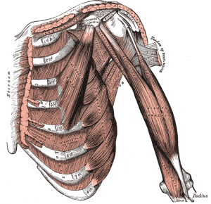
Learn more about how muscles work, what they look like, and how they're treated. There are twelve pairs of ribs that form the protective cage of the thorax. The first drawing showcases the latissimus dorsi muscles at the side of the ribcage. I think we have a respectable sense of how muscles contract on the molecular level let's take a step back now and just understand how muscles look at least structurally. Noticing the relationship of the latissimus and the teres ma развернуть. Muscles are tissues that contract to help parts of the body move. The skull, ribcage and pelvic bone are fairly solid and rigid parts of the body (though not always completely rigid). We've got this serratus anterior, which you can see laterally. Our ribcage exists to protect the heart and lungs. Visceral gross anatomy of a skeletal muscle most skeletal muscles are attached to two bones through tendons. Click now to learn more at kenhub! See more ideas about anatomy art, anatomy drawing, anatomy sketches. Anteriorly, they continue as cartilage, known as costal cartilage.
It is responsible for moving your ribcage towards the pelvis. This muscle is in the middle and has no muscles posterior to it. There are around 650 skeletal muscles within the typical human body. I think we have a respectable sense of how muscles contract on the molecular level let's take a step back now and just understand how muscles look at least structurally. This is a table of skeletal muscles of the human anatomy.
The Intercostal Muscles of the Ribcage from corewalking.com They also contract involuntarily, but have a striated appearance. Anatomy of a muscle cell. Our ribcage exists to protect the heart and lungs. Outside the ribcage, we've got three muscles really. We've got this serratus anterior, which you can see laterally. They are curved and flat bones. This muscle is in the middle and has no muscles posterior to it. They are further categorized according function such as flexion, extension, or rotation.
The first drawing showcases the latissimus dorsi muscles at the side of the ribcage.
When the ribcage is fixed contraction results in a posterior pelvic tilt. Spinoscapular and spinohumeral muscles, spinocostal muscles, modified intercostal musculature, trapezius muscle. They are curved and flat bones. Noticing the relationship of the latissimus and the teres ma развернуть. Cardiac muscles are found in the walls of the heart. I think we have a respectable sense of how muscles contract on the molecular level let's take a step back now and just understand how muscles look at least structurally. This muscle is in the middle and has no muscles posterior to it. They are further categorized according function such as flexion, extension, or rotation. The skull, ribcage and pelvic bone are fairly solid and rigid parts of the body (though not always completely rigid). This article covers the anatomy of the muscles of mastication, including their origins, insertions, and innervation. They're shapes that won't change too much between poses. Muscle types there are three types of muscle tissue: Each type of muscle tissue in the human body has a unique structure and a specific role.
Other muscles, like the skeletal muscle that moves the arm, is controlled by the somatic or voluntary nervous system anatomy rib cage. Muscle types there are three types of muscle tissue:

